Visual Electrophysiology Unit
The Visual Electrophysiology Unit (VEU) provides a comprehensive range of electrodiagnostic tests to detect, diagnose, and monitor retinal and/or visual pathway diseases and evaluate treatment interventions.
We perform a variety of tests that assess visual pathway function, from the retina to the occipital cortex. All the tests we perform adhere to and exceed International Society for Clinical Electrophysiology of Vision (ISCEV) standards. In addition to our electrophysiology testing, we perform a variety of colour-vision assessments to diagnose colour deficiencies. Our standard of care includes visual acuity test(s) including contrast vision and a standard color vision assessment. Additional tests or alternative tests may be performed to enhance patient care.

Electrodiagnostic Eye Testing
Our team is committed to providing exceptional patient care and high-quality visual electrophysiology testing; performing over 800 procedures on children of all ages every year.
The VEU is a referral-based testing cetre. We serve patients within the Department of Ophthalmology and Vision Sciences, including patients from community ophthalmologists and optometrists across Ontario and for some families across Canada and the USA.
We strive for a positive test-experience while doing our best to get reliable, quality results.

Programs and services
The VEU offers a variety of specialized tests. Our tests assess functional activity in the visual system using carefully planned visual stimuli (i.e., flashes, patterns). Multiple tests are often performed together to effectively evaluate different components of the visual pathway. Due to the specialized nature of our testing, some tests require more cooperation for a reliable interpretation. Test interpretations may take up to 2 months to complete and are sent to the referring physician for medical disclosure.
Check out the VEU’s programs and services below for more details.
An electroretinogram (ERG) measures the electrical response from the retina using a variety of flash stimuli that vary in intentisty, duration, and colour or to pattern stimuli that vary in size and frequency. An ERG requires the use of skin- and eye-electrodes for measrument. There are different types of ERGs.
Full-field ERG (ff-ERG)
- The full-field ERG is the most common test of global retinal function. An eye-electrode measures retinal function under dark- and light-adapted conditions to assess the function of rods and cones, respectively.
- Because the measurements are taken from the eye itself, this test can be performed awake (typically six years and older) or under sedation/general anesthesia.
- The test takes approximately 40 minutes. For accuracy in measurement, the pupils are dilated, and patients are encouraged not to blink or squint during the flashes. Contact lenses must be removed due to the use of eye-electrodes.
Multi-focal ERG (mfERG)
- The mfERG helps to assess macular cone function under normal lighting conditions, following pupil dilation. An eye-electrode measures the response from the cones to a large honeycomb pattern, where each honeycomb alternates between black/white during testing. This test measrues allows is to evalaute smaller areas of cone function, corresponding to a single honeycomb from the whole pattern.
- The test takes approximately 15 minutes. For accuracy in measurement, the pupils are dilated, and patients are required to not to blink or close their eyes while focusing on a target for each recording.
Pattern ERG (pERG)
- The PERG measures the overall electrical response from the central and peripheral macula. An eye-electrode measures the response from the cones and ganglion cells, while the eyes are stimulated by a checkerboard pattern at two distances (1 and 0.5 meters).
- The test takes approximately 20 minutes. For accuracy in measurement, glasses must be worn if used, and there should be minimal blinking and good focus. Contact lenses cannot be worn for this test.
Hand-held ERG
- The hand-held ERG is a quick screening tool to measure global retinal function and is useful in very young children or in children with co-morbidities that cannot undergo sedation/anesthesia. For this test, the eye-electrode is placed on the lower eye-lid to measure retinal function under dark- and light-adapted conditions. Unlike the ff-ERG, the hand-held ERG can only provide broad information on rod and cone system function - it is not as in-depth or sensitive as the ff-ERG, and each eye must be tested separately.
- The test takes approximately 1 hour. For any measurement to take place, the eyes must be open enough for the device to track the pupil.
The EOG assesses the function of the retinal pigment epithelium (RPE) during dark-adapation and light-adaptation. For this test, electrodes placed at the inner and outer corner of each eye. During testing, patients are required to make quick eye-movements (left to right) between two lights for 10 seconds, every minute in the dark (for 15 minutes) and then in the light (for 15 minutes).
The duration of the test itself is approximately 40 minutes. For accuracy in measurement, the pupils are dilated, the patient’s head must be very still and straight, and the eye movements must be quick between the two lights. Patients are encouraged not to squint, tilt their head, or make other eye movements during the recordings. All recordings must be obtained to evaluate RPE function.
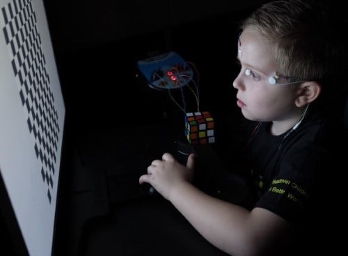
A VEP measures macualr and/or optic nerve/pathway function or visual processing, at the primary visual cortex (v1). It relies all parts of the visual pathway, from the front of the eye to the brain and requires your best corrected vision (i.e., glasses, if worn). For testing, skin-electrodes are placed on the forehead, top of the head, and on the back of the head. Each eye is tested together (binocualrly) and separately (with one eye covered).
There are multiple types of VEP protocols offered. These protocols are tailored to assess specific parts of the visual pathway based on patient ability, cooperation, and diangostic need.
- Flash – uses a bright flash to measure the ability to see light or visual potential.
- Pattern Reversal – uses checkerboard pattern of various sizes to measure macular and/or optic nerve/pathway function
- Pattern Onset-Offset – uses checkerboard pattern of various sizes and/or contrast to objectively measure visual function or evalaute visual potential
- Multi-channel – uses checkerboard and flash stimuli with multiple electrodes across the back of the scalp to assess for chiasmal misrouting.
The duration of these test ranges from 10 to 30 minutes. For accuracy in measurement, the patient must be awake, and focused on the stimulus. Glasses or contact lenses must be worn, if used. Atropine drops must be stopped 3 days prior to testing, if used for myopia control. Parental support and encouragement is required for infants or young children.
The FST uses patient-feedback to measure the dimmest perceivable light in patients with underlying retinal conditions; this test provides information on residual photorecetpor function in the most senstive region of the retina and is an outcome-measure for treatment interventions. Prior to testing, patients are dilated and dark-adapted for 40 minutes. During the test, patients must respond report if they 'see' and 'do not see' any flash of light with buttons, until a reliable minim threshold can be mesured. No electrodes are used for this test.
The duration of the test is approximately 1.5 hours. For accuracy in measurement, the patient is tested in complete darkness, must be alert at the time of testing, and respond to every stimulus. Each eye is tested at least twise to 3 wavelengths of light, to demonstate reliablity and evaluate photoreceptor function.
Color vision is the ability to see different wavelengths of light and helps us assess function of central retina and optic nerve/pathway. Different colour vision testing measures the type and extent of colour blindness in a patient.
There are 3 common types of colour vision tests used in the VEU.
Hardy-Rand and Ritter (HRR)
- A standard colour vision test that measures the degree of red-green and blue-yellow colour-vision.
- Patients are required to name the shapes that stands out on a page of dots and the colour of these shapes.
D-15 (Farnsworth and Lanthony)
- D15 tests require patients to arrange 15 coloured discs into a box starting from a specific colour. The discs have to be arranged so the colours blend from one disc to the next as smoothly as possible, like a rainbow.
- How the discs are arranged measures the degree of red-green, and blue-yellow colour vision.
Mollon-Reffin “Miminal” Vision Test
- Uses patient’s judgement of colours to measure the degree of red-green and blue-yellow colour-vision.
- Patients are required to pick out a stand-out colour disc from a group of discs. The lightest coloured disc that can be picked-out reveals your level of red-green and blue-yellow colour-vision. This is a fun and easy test to do in younger children.
Prepare for a visit to SickKids’ VEU
To prepare for a visit and for more information, learn more about each of our tests below.
If you’re a provider and need more information on VEU services, please contact us via email visualelectrophysiology.unit@sickkids.ca.
Volunteer opportunities
Interested in participating in testing, or being involved in visual electrophysiology? Contact us directly.
Fellowships and training opportunities
This is a one-year program offered through the University of Toronto and Visual Electrophysiology program. Prospective trainees are required to understand the principles of visual electrophysiology and develop skills in clinical interpretation. Weekly training sessions are included year-round to establish a thorough understanding of electrophysiology.
Two-week observerships are available for practicing ophthalmologists with an interest in electrophysiology and electrophysiologists. These observerships are tailored to individual needs. We will work with you to enhance your knowledge and build experience in visual electrophysiology testing and interpretation.
If you’re interested in events related to visual electrophysiology, visit the International Society for Clinical Electrophysiology of Vision site for dates in 2021.
Research
Current ongoing research within the VEU include research on:
- Electrophysiology testing in subjects undergoing gene therapy
- Identifying novel genotype-phenotype correlations in inherited rential dystrophies
- Implementing personalized testing protocols for Precision Child Health
Join Our Vision Research Study at SickKids!
We’re inviting kids (6+), adults, and seniors with healthy vision to take part in a one-time research visit at SickKids. Your participation will help advance eye health research and support children with inherited retinal diseases.
Here's what you can expect:
- Contribute to cutting-edge vision science
- One visit (up to 4 hours)
- Earn 5 volunteer hours
- Parking and travel costs reimbursed
Interested? Contact us at anupreet.tumber@sickkids.ca or 416-813-6133.
Our team
We are experienced visual electrophysiologists and members of the International Society for Clinical Electrophysiology of Vision (ISCEV). We are dedicated to providing individualized testing and the best clinical care.

- Ajoy Vincent – Medical director of the VEU & Staff Ophthalmologist (Ocular Genetics Program)
- Anupreet Tumber – Visual Electrophysiology Technologist, COA, M.Sc.
- Winnie Chou – Visual Electrophysiology Technician, RPN
Contact us
Email: visualelectrophysiology.unit@sickkids.ca
Phone: 416-813-6133
Location: Main Floor, Burton Wing, M293 (Eye Clinic)
Address
The Department of Ophthalmology and Vision Sciences
The Hospital for Sick Children (SickKids)
Main Floor, Burton Wing
555 University Avenue
Toronto, Ontario M5G 1X8

