Diagnostic Imaging

The Department of Diagnostic Imaging is dedicated to improving and enhancing patient care and the health of children. Our mission is to be a benchmark paediatric radiology site by embracing new technologies and utilizing state-of-the art equipment efficiently and effectively.
We provide an environment that supports and promotes clinical, academic and research excellence, and builds departmental clinical research expertise focused on the strengths of the adjacent clinical and research community.
What we do
The Department of Diagnostic Imaging (DI) employs a wide range of technologies in order to provide the highest standard of care for all our patients. Our department is subdivided into specialties: CT, GI/GU, Image-Guided Therapy, MEG, MRI, Nuclear Medicine, Ultrasound and X-Ray. Collaborating with the radiologists and technologists in these subdivisions as well as the dedicated staff who run Information Services, Research, Nursing and Quality and Safety contribute to making every patient's visit in DI run smoothly.
Programs and services
Expand each section below for more information, including individual contact details, photos and what to expect during your appointment.
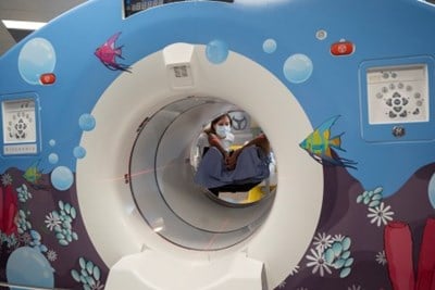
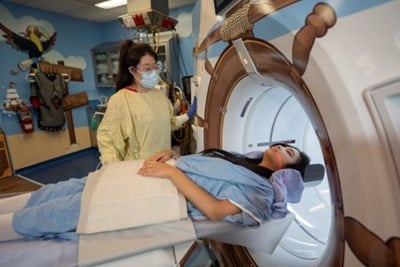
A CT (computed tomography) scan is an X-ray exam using special equipment and computers. The CT equipment takes a series of detailed X-ray pictures which provide a 2-dimensional image of the body but the data can also be used to construct 3-dimensional images. Sometimes, X-ray contrast is used to highlight specific organs, tissues and blood vessels. The X-ray contrast is given through an IV tube placed in a vein or the child might be asked to drink the X-ray contrast. This will depend on the type of procedure being performed.
Most CT exams are finished between 15 and 30 minutes depending upon the type of procedure being done, the severity of illness and the age and co-operation of the child. The CT scanner is a large machine with a hole in the center and some of our patients say it looks like a large doughnut. Your child will lie on the table that moves through the donut hole. CT scans are fast and painless, but it is very important that your child holds still on the table for long enough to complete the test. Any movement will cause the images to be blurry. The CT machine will not touch your child.

A nurse from Diagnostic Imaging will review the CT orders and contact you if necessary to determine if your child needs sedation or general anesthetic for the CT scan.
Our radiologists and technologists are specialists in pediatric imaging. In all CT procedures we strive to minimize the amount of ionizing radiation applied, using only what is necessary to achieve optimal imaging results. To learn more about Radiation Safety at SickKids please see our website.
Parents that are pregnant or may be pregnant will not be allowed to stay in the x-ray room with their child. Therefore, please make sure to bring someone else with you who can stay with your child during the test, if needed.
The CT images will be reported by a pediatric radiologist and the results will be sent to the physician who ordered the procedure.
Contact
Manager: Michelle Cote
Team Leader: Sandi Sutton
SickKids has one PET/CT scanner in the department of Diagnostic Imaging which provides incredibly detailed images by fusing two state of the art technologies.
While the PET portion allows us to look at metabolic function by monitoring glucose metabolism in the body, the CT portion provides detailed anatomical information of various organs in the body. These combined images provide our referring physicians with comprehensive information that helps them make necessary treatment recommendations and/or alterations to existing treatment plans for patients. PET/CT studies are performed using a camera capable of acquiring both CT data and PET data while the patient remains on the same imaging bed.
The scan room has been designed to provide a child friendly environment to help make this scan as comfortable and easy as possible. One parent can remain in the room during the scan if necessary to help reassure their child.
PET/CT scans can take anywhere from 10-60 minutes to complete depending on the area being scanned and require your child to remain still for the entire time. Your child will be asked to have nothing to eat or drink for 4 hours prior to their appointment time in order to get high quality diagnostic images. Someone from Sick Kids will contact you prior to your appointment to confirm preparation details.
Contact and location
The PET/CT scanner is located on the 2nd Floor of the Burton Wing and can reached by calling 416-813-6065.
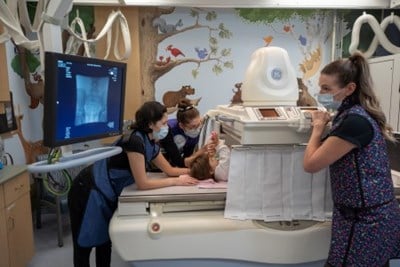
General Radiography, commonly referred to as “x-ray” is an essential diagnostic tool and was the first established modality of Diagnostic Imaging.
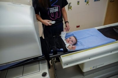
When an x-ray is taken, a very small amount of radiation is used to create a two-dimensional view of the inside of the body. The technologist will usually take two to three images from different angles of your child in different positions to provide the best quality images for the radiologist to report. These images are used to detect abnormalities in bones, lungs and other internal organs.
Duration times for x-ray exams range from 10 minutes to 90 minutes depending upon the number of views required, the severity of illness and the age and co-operation of the child. The x-ray machine will not touch your child. Just like in photography, it is very important that your child holds still during the “picture” as any movement will cause the images to be blurry.

Your child will not feel the x-rays, but sometimes the positions needed to get the best images are awkward or uncomfortable. Our certified technologists are experts in working with children and will make your child as comfortable as possible during the exam.
Parents that are pregnant or may be pregnant will not be allowed to stay in the x-ray room with their child. Therefore, please make sure to bring someone else with you who can stay with your child during the test, if needed.
High quality imaging at a very low radiation dose is our primary focus. The x-rays will be read by a pediatric radiologist and the results will be sent to the physician who ordered the x-ray.
Contact
Hours of Operation: Monday to Friday, 8 a.m. to 4 p.m.
There are no outpatient appointments or walk-ins on weekends. Emergency and inpatient service only.
Download our Requisition Form (PDF)
Modality Head: David Manson
Manager: Michelle Cote
Team Leader: Sandi Sutton
Main Contact:
Phone: 416-813-4960
Fax: 416-813-7644
Location: Main Floor Atrium (beside Starbucks)
In GI/GU x-ray, we visualize disorders of the Gastro-Intestinal or Genito-Urinary system. We use a combination of fluoroscopy and X-ray contrast. Fluoroscopy is an imaging technique that uses X-rays to create “real time” or moving images of the body. X-ray contrast is used to highlight specific organs so we can see the area being studied.
Our radiologists and technologists are specialists in pediatric imaging. In all GI/GU x-ray procedures we strive to minimize the amount of ionizing radiation applied, using only what is necessary to achieve optimal imaging results. To learn more about Radiation Safety at SickKids, please see the section below on Diagnostic Imaging (DI) Quality and Safety.
Some of our procedures may be completed quickly within 30 minutes while some can continue for several hours. X-ray contrast can be given in various ways depending on the study. To look at swallowing, or the GI tract (esophagus, stomach, small intestine, large intestine), the patient will drink the X-ray contrast. To look at the urinary system (bladder, ureters, kidneys, urethra), the X-ray contrast will be put into the bladder with a catheter. To examine the large intestine or rectum, X-ray contrast may flow into the patient using an enema tube.
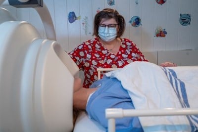
The GI/GU rooms have a flat table with a camera that pulls over the patient to create a small tent. The radiologist or technologist will move the camera up and down to best see the area being examined. The room light will dim to allow the images to be seen with more detail. During the test, the machine might make some noises and we may tilt the table up and down. One person is allowed in the room with the patient during the study, but siblings under the age of 18 are not allowed to stay with the parent and patient during imaging.
Parents that are pregnant or may be pregnant will not be allowed to stay in the x-ray room with their child. Therefore, please make sure to bring someone else with you who can stay with your child during the test, if needed.
The x-rays will be reported by a pediatric radiologist and the results will be sent to the physician who ordered the procedure.
Contact
Phone: 416-813-6068
Fax: 416-813-6043
Location: 2nd floor, Burton Wing (follow the signs for Diagnostic Imaging)
Modality Head: Ailish Coblentz
Manager: Michelle Cote
Team Leader: Sandi Sutton
Image-Guided Therapy (IGT) is a state-of-the-art facility that was specially designed to house the growing interventional radiology service, and to facilitate many other disciplines to work together to perform image-guided treatments. Several procedures can now be performed safely in one place, on the same day, under one anaesthetic often resulting in a shorter hospital stay to the benefit of the patient.
The IGT division is headed by Dr. Joao Amaral, and is part of the Department of Diagnostic Imaging (DI), under the leadership of the Radiologist-in-Chief, Dr. Manohar Shroff and the Director of Diagnostic Imaging Wayne Lee.
IGT is a member of the Centre for Image-Guided Care (CIGC) and is utilized as the main platform for delivering clinical care through this Centre.
About us
The IGT Clinic provides pre-procedure planning and communication with the child and their family. Families can discuss the details of a procedure with IGT staff to make an informed decision about their child’s care. Positive outcomes from the consultation include optimizing quality, efficiency and thorough patient care with improved workflow.
Contact us
Julie Zettel, Clinic Nurse
Phone: 416-813-7654 ext. 201804
Email: julie.zettel@sickkids.ca
Image-Guided Therapy (IGT) Requisition Form
Clinic/registration location
The IGT Clinic is located on the second floor of the Atrium. Take the Atrium elevators to the second floor and look for the IGT sign, which will lead you to the IGT registration desk.
Once registered, a SickKids identification (ID) band will be placed on your child. The band must be worn on your child’s arm or ankle at all times. Check your child’s ID band to make sure the information is right. If your child is not wearing a band or has the wrong one, it can lead to errors that can result in the wrong care for your child. Staff must always check the band before providing care to your child. Your child must keep the band on at all times while at SickKids.
Vascular Anomalies and IGT
Many children with vascular malformations are treated by interventional radiological procedures, which are carried out in IGT. Our interventional radiologists from IGT lead the Vascular Anomalies Clinic team. They perform the important interventional radiological procedures such as sclerotherapy, embolization and endovenous laser needed to treat some patients with vascular malformations. Many patients with vascular malformations will benefit from these minimally invasive treatments.
Magnetoencephalography (MEG) is a special machine that is much different from an MRI in both function and appearance. We have been performing studies in our lab for over twenty years. MEG non-invasively measures magnetic fields created by the brain’s electrical activity (fields that are as low as pico-Tesla). MEG is used primarily to map epileptogenic foci and eloquent brain function. A special MRI sequence is completed as part of a MEG study. The MEG data is mapped onto the MRI images and can show the correlation between epilepsy, function and brain anatomy.
Children with epilepsy are the primary patient group benefiting from this technology at present. MEG is utilized to determine surgical candidacy for patients that have refractory seizures. MEG is used primarily to map interictal seizure activity. In addition, MEG also provides functional information used for surgical planning in neurosurgery patients with conditions other than epilepsy.
Analysis of MEG data does take time and especially for epilepsy assessment, all clinical cases must be approved through the Neurophysiology department prior to booking. Only requests from Neurologists or Neurosurgeons will be accepted for MEG.
Magnetoencephalography (MEG) is a joint venture between the Epilepsy Service of Neurology and the Department of Diagnostic Imaging. The Peter Anderson Clinical MEG is located within the clinical MRI department on the service floor of the hospital (Room S763). For epilepsy mapping MEG is used to localize between seizure or interictal activity. Eloquent brain functions can also be mapped such as somatosensory response, visual response, auditory response and language.
Magnetic Resonance Imaging (MRI) produces images through the use of a strong magnetic field. MRI can be used to visualize internal anatomy to assist in diagnosis and treatment planning.
During a MRI exam, patients need to remain still usually between 30min to 90min depending on the type of exam and resulting images. In some cases, children require sedation or a general anesthetic for their appointment.
In order to assist children during their MRI exam, our expert team of technologist and nurses will foster a friendly and inviting environment for your child. Your child will have time to observe the MRI machine and feel comfortable with all its components. Depending on the type of exam, your child may be eligible to watch a movie during the exam process with the help of specialized googles and headphones. Parents are encouraged to bring their child’s favorite DVD along with them to their MRI appointment.
For MRI exams not requiring a general anesthetic, a parent may accompany their child into the MRI room for the exam. They can sit beside their child to help them feel safe and comfortable during the MRI process.
Depending on your child’s age, you may be eligible for a “Feed & Sleep” exam as an alternative to a general anesthetic. Typically, babies are most successful with this exam type. It involves keeping your child hungry and active for several hours before the MRI appointment time. When you arrive in the department the technologist will instruct you to feed your child and help them fall asleep. Afterwards, a parent will carry their child into the MRI room where the technologist will position the patient and begin the exam.

MRI Safety
MRI safety is our number one concern at SickKids. Patients who are coming in for an MRI exam must successfully undergo an MRI safety screening process performed by authorized MRI personnel. On arrival at the MRI department, the patient or their parent will be asked to complete and date a patient MRI screening form. Parents will be notified of any concerns. If the patient is approved for the exam, they will be given a set of hospital clothing to change into before going beyond the reception area. At this time, all metallic objects such as jewelry, watches, hair pins and rings must be removed. Before the patient enters the MRI room, all sections of the patient MRI screening form will be reviewed by MRI personnel.
We recommend parents review the patient screening form before their appointment. It is important to know all implants in your child before you come for your appointment. For implants that are not from SickKids please ensure you have the make and model of the implant, this will minimize delay during your appointment. This information can be obtained from the hospital or office the implant was from. Please call MRI reception if you have any questions or concerns.
Parents who wish to accompany their children during the MRI procedure must be properly screened and educated to avoid injury. Parents must undergo a MRI screening process performed by MRI personnel and complete a non-patient MRI screening form before they enter the MRI room. They will also need to remove all metallic objects such as jewelry, watches, hair pins and rings.
Both patient and accompanying parent will be given hearing protection during the MRI exam. Staff at SickKids ensure that equipment, technology and procedures are tailored to meet the unique needs of our pediatric patients.
For more information on what you can expect on your visit for MRI exams, please visit AboutKidsHealth.ca, see the MRI information page. If your child is scheduled for a Magnetic Resonance Enterography exam (MRE) please watch the video of a virtual MRE tour to have a better idea of what to expect.
Referring physicians
Thank you for referring your patient to our MRI Department.
MRI History and Physical Form (PDF)
MRI Patient Screening Form (PDF)
MRI Contrast Screening Form (PDF)
SickKids offers two types of MR Scanners:
- 3 Tesla (3T) MRI Scanner
- 1.5 Tesla (1.5T) MRI Scanner
Patient Safety and timeliness are our utmost priorities. You will ensure that your patient is scheduled appropriately by accurately completing the request for consultation included below. Patient bookings are done according to information you provided on the MRI requisition. In order to assist you in that process, below are some guidelines.
Guidelines for completing an MRI requisition:
- Include the patient’s weight and height. They assist us in proper booking and planning
- Please evaluate the ability of the patient you are referring us to be able to hold still for about an hour. If you feel this is not possible, we may need to sedate or anesthetize the patient. Your clinical evaluation will allow us to appropriately schedule the procedure in a timely manner. As a rule of thumb patients above six years of age usually are able to undergo their MRI exam without being sedated
- List all surgeries—please indicate the procedure and date of surgery. Please include minor surgeries such as dental interventions or ear tube implants
- Please document all Implants- Implants which are MR Safe on the 1.5T magnet may not be safe on the 3T magnet
- Name of Implant, and date implanted
- Manufacturer of Implant
- Model number of Implant
- Indicate if there are any renal issues, or if there has been a prior contrast reaction. It is general practice to calculate the GFR on all patients who need MRI contrast if there is suspicion of compromised renal function.
Contact
MRI Reception: 416-813-1500 Ext. 203546
Address:
SickKids Service Level
555 University Ave.
Toronto, ON
M5G 1X8
MRI Team Leader: Joey Cheng-Singleton
MRI Quality Lead: Jue Hou
MRI Flow Coordinators: Imran Arif, Leslie Burns, Andrea Young
MRI Safety Officer: Stephanie Holowka
Clinical Nurse Specialist: Ingrid Costa
MRI Manager: Nicole Bennett
Modality Heads (Body): Mary-Louse Greer, Govind Chavhan
Modality Head (Neuro): Birgit Ertl-Wagner
Hours of Operation
Monday to Friday - 7 a.m. to 10 p.m.
Saturday and Sunday - 7 a.m. to 3 p.m.
Long weekends and holidays, only on call staff are available. There is no one at reception.
What is Nuclear Medicine?
Nuclear Medicine (NM) is a specialized area of radiology that uses very small amounts of radioactive materials (radiopharmaceuticals) to examine organ function and structure and is a valuable tool in the diagnosis and treatment of a variety of illnesses and medical conditions. Tests in nuclear medicine are often done in multiple parts and can take anywhere from 30 minutes to several hours to complete. Occasionally you may be asked to return over a period of several days but this will be communicated to you when your test is scheduled.
You can find more information about Nuclear medicine tests on AboutKidsHealth.
How is it done?
The radiopharmaceuticals are given primarily by intravenous injection although for some studies it may be given by inhalation (breathing) or ingestion (eating). This is done by a registered Nuclear Medicine Technologist (MRT(N)) and every effort is made to make this process as easy and painless as possible for the patient. The use of topical anesthetic cream (maxilene), lidocaine spray to temporarily numb the skin, sucrose (a mild analgesic which is effective in decreasing short-term pain and distress during minor procedures for neonates and infants), distraction techniques (watching something on an iPad, phone ) and breathing techniques are effective methods to help reduce anxiety and discomfort.

The radioactive substance is absorbed by body tissue and gives off radiation (gamma radiation) which is detected by a specialized piece of equipment known as a gamma camera. The gamma camera may remain stationary resulting images that are two-dimensional (2D) or it may rotate around the patient to create three dimensional (3D) images similar to those obtained by a CT scan. This is known as Single photon emission computed tomography, or SPECT. SickKids currently has 3 gamma cameras and a PET/CT scanner which allows us to look at function and anatomy simultaneously.
The division of Nuclear Medicine also provides Bone Mineral Density (BMD or DXA) imaging. This test uses a low dose x-ray to look at the strength and health of bones and is particularly helpful in the diagnosis of osteoporosis and osteopenia. This test does not require the injection of a radiopharmaceutical and usually takes 15 to 30 minutes.
Where is Nuclear Medicine located in the hospital?
We are located on the 2nd floor of the Burton Wing. You will need to follow the signs for Diagnostic Imaging and check in at the registration desk approximately 15 minutes prior to your scheduled appointment time.
Contact
The Nuclear Medicine department is open Monday to Friday - 8 a.m. to 4 p.m.
Our information coordinator can be reached by calling 416-813-6065 if you have any questions regarding your or your child’s appointment.
Division Head: Dr. Amer Shammas
Manager: Nicole Bennett
Team Leader: Nancy Ribeiro
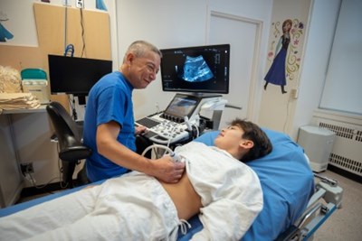
Ultrasound is a safe, non-invasive and inexpensive test done as a routine primary examination for all pediatric patients. It works with high frequency soundwaves and needs minimal preparation or no pre examination preparation in most cases. Almost all internal organs can be examined with ultrasound. This test is painless and has no ionizing radiation.
As one of the largest paediatric sonography centres in North America, our team of ultrasonographers, radiologists and support staff are dedicated to the provision of excellent clinical service.
Modality Head: Ricardo Faingold
Manager: Nicole Bennett
Team Leader: Jennifer Whitmore
The Department of Diagnostic Imaging (DI) relentlessly pursues innovative solutions to meet the needs of each and every patient and achieve the highest quality of patient care. We are committed to the provision of exceptional services while recognizing and respecting the diversity of the patients and other customers we serve.
We take a systematic approach to visualize the future in the context of internal and external business environment, build organizational capacity for innovation and formulate strategies to overcome challenges and maximize future opportunities. Comprehensive strategies are developed to continually improve quality of care, enable equitable and timely access to services, further enhance patient safety, introduce advanced technology and equipment, create value for patients and their families, and promote clinical, academic and research excellence.
We employ a full-time medical physicist who monitors and minimizes radiation doses, and have established an MR Safety Committee who oversees and ensures the safety of our patients.
Contact
Quality Manager: Laura Belsito, Senior Manager of Operations
Medical Physicist: Nicholas Shkumat
MR Safety Officer: Stephanie Holowka
The Diagnostic Imaging Information Services Division was established in 1999 as our department moved from a film to a filmless environment. It is responsible for maintaining and enhancing all the major computer applications within Diagnostic Imaging. These applications include the Picture Archiving and Communication System (PACS), the departmental Radiology Information System (RIS), Voice Recognition (VR), and all associated hardware and software.
CD Uploading Protocol
DI Electronic Imaging Department (Film Library)
For information about CD/DVD, Secure FTP and DI Reports, please contact us at 416-813-7511, Monday to Friday, from 8 a.m. to 5 p.m.
PACS Support: Monday to Friday, from 7 a.m. to 5:30 p.m.
Phone: 416-813-8358
PACS urgent requests: 416-713-0187
Manager: Fatima Lima-Simao
Telephone: 416-813-4965
Nursing in DI requires a set of advanced skills and as nurses working within a high tech environment, there is the opportunity to promote and to provide quality-nursing care to children.
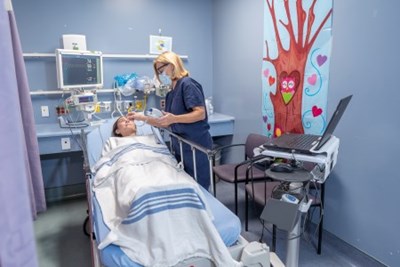
The nurses utilize physical assessment skills, perform guided risk assessments, and determine a sedation plan taking into consideration all potential risks to the patient. The care provided by our nursing team spans from initial assessment, to induction, monitoring and recovery.
Nurse Educator: Darlene Murray
Clinical Nursing Coordinator: Wendy Padilla
Who we are
The Department of Diagnostic Imaging is dedicated to improving and enhancing patient care and the health of children. Our mission is to be a benchmark paediatric radiology site by embracing new technologies and utilizing state-of-the art equipment efficiently and effectively. We provide an environment that supports and promotes clinical, academic and research excellence, and builds departmental clinical research expertise focused on the strengths of the adjacent clinical and research community.
Radiologist-in-Chief: Dr. Birgit Ertl-Wagner
Director: Christine Hill
A list of our key staff can be found below, with links to their email and/or SickKids Staff Profile.
Laura Belsito – Senior Manager, Diagnostic Imaging Operations
Michelle Cote – Senior Clinical Manager, CT/GIGU/IGT/X-ray
Nicole Bennett – Senior Clinical Manager, MRI, Nuclear Medicine, Ultrasound
Fatima Lima-Simao – Manager, PACS/RIS, Patient Coordinator and Clerks
- Darlene Murray - Nurse Educator
- Denise Carraretto - Clinical Instructor: Radiography
- Jose Jarin - Clinical Instructor: Ultrasound
- Mandy Kohli - Clinical Instructor: Nuclear Medicine
- Evan Kitamura – IGT
- Nancy Ribeiro – Nuclear Medicine
- Jennifer Whitmore – Ultrasound
- Sandi Sutton – Atrium X-ray, GI/GU X-ray and CT
- Joey Cheng-Singleton – MRI
- Stephanie Holowka – MEG, MRI Safety Officer, Computer Navigation, and 3D Imaging
Radiologists
- Dr. Joao Amaral
- Dr. Helen Branson
- Dr. Govind Chavhan
- Dr. Ailish Coblentz
- Dr. Alan Daneman
- Dr. Andrea Doria
- Dr. Birgit Ertl-Wagner Radiologist-in-Chief
- Dr. Ricardo Faingold
- Dr. Alessandro Gasparetto
- Dr. Mary-Louise Greer
- Dr. George Koshy Chiramel
- Dr. Pradeep Krishnan
- Dr. Christopher Lam
- Dr. Suzanne Laughlin
- Dr. Cathy MacDonald
- Dr. David Manson
- Dr. Claudia Martinez-Rios
- Dr. Elka Miller
- Dr. Prakash Muthusami
- Dr. Oscar Navarro
- Dr. Kamaldine Oudjhane
- Dr. Dimitri Parra
- Dr. Mike Seed
- Dr. Amer Shammas
- Dr. Manohar Shroff
- Dr. Jennifer Stimec
- Dr. Jeffrey Traubici
- Dr. Reza Vali
- Dr. Shi-Joon Yoo
Scientists (affiliated with the SickKids Research Institute)

One of the goals of the Department of Diagnostic Imaging (DI) is to promote research excellence. Our DI research group was initiated to exploit rapidly emerging technologies for diagnostic procedures for children.
Our department supports active research and training programs for undergraduate and graduate students, medical students, residents and post-doctoral fellows. Our fellows, radiologist and scientific staff undertake research activities within our multiple imaging modalities. We also support research projects emanating from outside our department and contribute to major research interests within the hospital and the Research Institute (such as Brain and Behaviour, and the Cardiovascular program).

The Department of Diagnostic Imaging sponsors four programs in medical education.
Our Medical Electives Program offers 4th year medical students from Canada and around the world the opportunity to work in our Department for two to four weeks. Our Residency Program offers up to a four-month rotation in pediatric radiology for residents at either the University of Toronto, Queen's University or McMaster University. The Fellowship Program sponsors graduates of radiology residency programs from Canada and around the world for a 1-2 year position within our Department in a specialized modality of Radiology. And finally, our Observership Program grants foreign trained medical professionals permission to observe specific procedures within Diagnostic Imaging to gain experience. Through our education programs, the Department of Diagnostic Imaging hopes to produce the next generation of health care professionals.
In the next section, you'll find detailed information about each of these educational offerings, including application processes.
Paediatric Radiology Fellowship Program
Dr. Oscar Navarro
Paediatric Radiology Program Director
The Clinical Fellowship Program in the Department of Diagnostic Imaging at The Hospital for Sick Children provides training for those radiologists interested in pursuing a career in paediatric imaging. The candidates must have already completed a four-year training in a diagnostic radiology residency program or must be board-certified as radiologists in their country of origin and need to meet the minimum eligibility requirements for licensure with the College of Physicians and Surgeons of Ontario. The program is within one of the largest and busiest paediatric radiology departments in the world, performing approximately 130,000 examinations per year, allowing exposure to practically the entire spectrum of normal and pathologic imaging findings one can encounter in paediatric practice. The Clinical Fellowship Program is open to both Canadian and international applicants and in fact as of April 2023 we have fellows from Canada, Costa Rica, Panama, Chile, Brazil, United Kingdom, France, Switzerland, Spain, Nigeria, South Africa, Saudi Arabia, India and Australia.
The Clinical Fellowship Program in our Department is part of the Fellowship Program of the Department of Medical Imaging of the University of Toronto. Our Program offers fellowships in Paediatric General Radiology, Paediatric Neuroradiology, Paediatric Interventional Radiology, Paediatric Cardiac Imaging, and Paediatric Nuclear Medicine. We have a variable number of funded and self-funded positions available, starting in July and sometimes also in January of each year. The funded positions are paid for by our Department whereas the funding of the self-funded positions should be arranged by the candidate either through international grants or sponsoring institutions in their home countries.
Most of our fellowships are one-year duration. In Paediatric General Radiology there is some flexibility and interested candidates may apply to one-year or two-year programs depending on the particular needs of the applicant or of the institution that is sponsoring the candidate. At the time of the application, the candidate can apply to more than one fellowship program, for example, one year of Paediatric General Radiology and one year of Paediatric Neuroradiology. However, each application is treated separately by the Fellowship Committee and most of the times the applicant will only be accepted at only one fellowship with recommendation to apply to the other fellowship by the next year.
Paediatric General Radiology Fellowship
The clinical fellowship in Paediatric General Radiology includes rotations in the following areas: plain radiographs (including in-patient studies from the pediatric and surgical wards and from the neonatal and paediatric intensive care units, as well as outpatient and emergency room examinations), fluoroscopy (gastrointestinal and genitourinary), ultrasound (including neuro-sonography), body CT, body MRI (chest, abdomen and musculoskeletal), neuroradiology (including CT and MRI), and nuclear medicine. Fellows have protected time to attend multiple one-hour teaching sessions every week and periodically participate at clinical case rounds in general surgery, oncology, emergency, non-accidental injury and gastroenterology.
Our training program in Paediatric General Radiology is currently accredited by the Royal College of Physicians and Surgeons of Canada (RCPSC) as a one-year Paediatric Radiology Residency Program. This means that those trainees that have completed a diagnostic radiology residency program in Canada and/or are certified by the RCPSC in Diagnostic Radiology and enter our program in Paediatric General Radiology will be recognized as PGY6 paediatric radiology residents. These trainees may be eligible for certification with examination in Pediatric Radiology by the RCPSC following successful completion of our program. Fellows that are not part of the RCPSC track may also take the RCPSC examination in paediatric radiology through the Subspecialty Examination Affiliate Program (SEAP) and become a subspecialist affiliate with the RCPSC.
In July 2024, our clinical fellowship in Paediatric General Radiology will start the implementation of Competence by Design (CBD), a specific model of competence-based medical education supported by the RCPSC. This is characterized by 4 well-defined stages of training, clear learning objectives called entrustable professional activities (EPAs), observation of trainees completing EPAS, documentation of observations in an electronic portfolio, and regular reviews of documented observations by the Competence Committee that will provide recommendations for progression to the next stage of training. Benefits of CBD include clear learning expectations for the trainees, more opportunities for feedback and coaching, more control of the learning by the trainees, preventing gaps in knowledge, and promotion of feedback in lifelong learning.
Read about the goals and objectives for the General Radiology Fellowship (PDF)
Paediatric Neuroradiology Fellowship
The clinical fellowship in Paediatric Neuroradiology provides training in diagnostic pediatric neuroradiology, including CT and MRI of the brain, head and neck, and spine, and covers a wide range of congenital and acquired conditions. Fellows gain competence in protocoling, monitoring and reporting CT and MRI studies from the fetus through to the age of 18 years including neonates. Multiple teaching and clinical rounds are held every week. Fellows are actively involved in the presentation of clinical case rounds and in the preparation of lectures and presentations for meetings. Fellows become part of the neuroradiology team and take part in on-call services in pediatric neuroradiology. Fellows actively interact with many clinical services especially neurosurgery, neurology, and otolaryngology/head & neck surgery.
Read about the goals and objectives for the Neuroradiology Fellowship (PDF)
Paediatric Interventional Radiology Fellowship
The clinical fellowship in Paediatric Interventional Radiology provides training for those radiologists with previous training in pediatric radiology or adult interventional radiology interested in pursuing a career in image-guided pediatric interventions. Our Division of Paediatric Interventional Radiology is one of the largest and busiest in the world, performing a wide variety of procedures, including but not limited to central venous access, biopsies, gastrointestinal access, management of post-surgical and post-transplant complications, angiography, and management of vascular anomalies. Fellows attend weekly theorical lectures on the above mentioned topics. Fellows are exposed and actively take part in numerous procedures under the supervision of staff interventional radiologists and it is expected that at the end of the fellowship they will be the main operators in the majority of interventions. Fellows are also actively involved in morning huddles, clinical rounds, clinic consults, and post-procedure patient management as well as in house, national and international conferences presentations.
Read about the goals and objectives for the Interventional Radiology Fellowship (PDF)
Paediatric Cardiac Imaging Fellowship
The clinical fellowship in Paediatric Cardiac Imaging provides training in MRI of congenital and acquired cardiovascular disease in children. With the recent acquisition of new CT equipment in our department, we expect that the training will also extend into cardiac CT. The program is a combined program for radiology and cardiology fellows. Fellows gain competence in designing, protocoling, monitoring, post-processing and reporting MRI. Importantly, the program is set in such a way that fellows work closely together with technologists to acquire basic knowledge and skills on MRI. In addition, radiology-based fellows learn basic cardiology concepts through daily interaction with colleague cardiology-based fellows. Fellows are also provided with ample opportunities to participate in clinical discussions and rounds, to attend a number of lectures organized by the Department of Diagnostic Imaging and the Division of Cardiology, Department of Paediatrics. Fellows are actively involved in research projects and presentations at scientific meetings.
Read more about the goals and objectives for the Cardiac Imaging Fellowship (PDF)
Paediatric Nuclear Medicine Fellowship
The clinical fellowship in Paediatric Nuclear Medicine provides training for board-certified nuclear medicine physicians or radiologists that are interested in paediatric nuclear medicine. The program enables fellows to learn a wide variety of nuclear medicine imaging techniques and procedures in children including PET/CT and SPECT imaging as well as radionuclide therapy. Upon completion of training, the fellow is expected to be competent in performing and interpreting common paediatric nuclear medicine examinations. Fellows are actively involved in teaching and participation in clinical rounds.
Read more about the goals and objectives for the Nuclear Medicine Fellowship (PDF)
General information
Fellows are trained and evaluated in all aspects of the CanMEDS competency framework (medical expert, communicator, collaborator, leader, health advocate, scholar and professional roles). Fellows gain competence through a process of graded responsibility by which they take on increased responsibility as they acquire greater experience under ongoing supervision of staff radiologists. This is supplemented by daily teaching sessions in all areas of paediatric imaging, participation in clinical rounds, and presentation at quality assurance rounds and journal club. Fellows are also expected to cover MRI evening shifts and to do after hours and weekend shifts on a rotating basis with the other fellows. As the fellows gain more familiarity with paediatric imaging and with the department, they are put in a more supervisory role over diagnostic radiology residents who are performing “in house” call during their paediatric imaging rotation in our hospital. Fellows are expected to reach the level of junior staff colleagues before they leave the department.
Protected academic time is provided to the fellows so they can work in research or quality improvement projects and to prepare presentations during the year. The fellows are supported in all academic activities and in the past, this has led to numerous presentations at national and international paediatric imaging meetings as well as to the publication of scientific papers, educational exhibits, and book chapters. Fellows are expected to be productive and in fact the Department of Medical Imaging of the University of Toronto requires production of at least one original research article or completion of an approved quality improvement project per year of fellowship in order to issue a certificate of completion of fellowship.
Paid vacation is 20 working days per fellow, per year. Additional time is provided to all fellows to attend conferences and/or courses. Some funding is available for travel and accommodation to one conference for each year of fellowship.
Goals and objectives
- Paediatric General Radiology (PDF)
- Paediatric Neuroradiology (PDF)
- Paediatric Cardiac Imaging (PDF)
- Paediatric Interventional (PDF)
- Paediatric Nuclear Medicine (PDF)
Fellowship applications
Following the current recommendations by the Society of Chairs of Academic Radiology Departments (SCARD) and supported by the Society for Pediatric Radiology (SPR), we are postponing the application process that we had initially stated on our previous posting. This applies to fellowships starting in January or July 2025.
- For international applicants, explicitly those that are not enrolled in an ACGME- or RCPSC-accredited program, the application process will open on October 1, 2023 and will close on November 10, 2023.
- For applicants that at the time of the application are enrolled in an ACGME- or RCPSC-accredited program, the application process will open on November 1, 2023 and will close on November 10, 2023.
The application process to all of the fellowship programs (Paediatric General Radiology, Paediatric Interventional Radiology, Paediatric Neuroradiology, Paediatric Cardiac Imaging and Paediatric Nuclear Medicine) is similar, including funded and self-funded positions.
In the application form there is updated information regarding available funded and self-funded fellowship positions at the time of the application. A completed application form, curriculum vitae, statement letter and three recent reference letters should be submitted by email before the deadline. All reference letters should be dated in 2023 and addressed to Dr. Oscar Navarro, Paediatric Radiology Program Director. In the preparation of the curriculum vitae, the candidate should clearly identify publications (peer-reviewed and non-peer-reviewed), oral presentations, posters and research work still in progress. All published work should be listed following standard format:
Grealish J, Haaland E, De Bruyne K. Effect of high intensity sport activities in the metaphyses of long bones in prepubescent children. J Pediatr Sports Med 2022; 65:731-738.
The candidate’s name should be in bold font. Always use the standard abbreviation of a journal’s name according to the ISSN List of Title Word Abbreviations (see ISSN.org LTWA).
Candidates can apply to two different fellowships (i.e. Paediatric General Radiology and Paediatric Neuroradiology, etc) using one single form. The Fellowship Committee will review all applications within 4-8 weeks after the deadline and will select a small number of candidates that will be called for an interview. We plan to hold remote interviews using a videoconference platform during the month of January 2024. The final decision about acceptance will be made within a few weeks after the interview.
It is expected that all fellowship candidates have advanced English language proficiency, both oral and written. Although we do not require that the applicant pass an English language exam, the candidate’s fluency in English will be evaluated at the time of the interview.
Orientation handbook for new residents and fellows
The 2022 Orientation Handbook for New Residents and Fellows at the University of Toronto is currently available and we expect the 2023 version will become available in July 2023. This downloadable booklet offers up-to-date information about topics such as registration, health care coverage, cost of living, accommodation, and child care and schooling, to help new learners, especially those from outside Canada, achieve a successful transition to residency and fellowship training at the University of Toronto.
Fellowship Application Form
Paediatric Imaging Fellowship Application (.docx)
Contact
For more information regarding Paediatric Radiology fellowships or the application process, please contact:
Diagnostic Imaging Education CoordinatorFellowship Program, Applications Process
Department of Diagnostic Imaging
The Hospital for Sick Children
555 University Avenue
Toronto, Ontario M5G 1X8
email: di.educationcoordinator@sickkids.ca
Audrey Onugha
Education Coordinator
Paediatric Radiology Program Administrator
Department of Diagnostic Imaging
The Hospital for Sick Children
555 University Avenue
Toronto, Ontario M5G 1X8
email: di.educationcoordinator@sickkids.ca
Oscar Navarro, MD
Fellowship Program Director
Department of Diagnostic Imaging
The Hospital for Sick Children
555 University Avenue
Toronto, Ontario M5G 1X8
email: oscar.navarro@sickkids.ca
Paediatric Imaging Rotation for Diagnostic Radiology Residents
Radiology Residents are an integral part of the Department. Residents from the University of Toronto, McMaster University and Queen's University rotate in Diagnostic Imaging for 3-4 months, as part of their overall residency program in radiology. The purpose of this policy is to ensure optimum supervision, instruction and evaluation for Radiology Residents in the performance and reporting of radiological procedures.
Application procedure
To complete a rotation in the Department of Diagnostic Imaging at The Hospital for Sick Children, students must be accepted to a residency program at the University of Toronto, Queen's University or McMaster University. Please contact your university for more information on rotations.
Paediatric Radiology Medical Electives Program
The Medical Electives program run by the Department of Diagnostic Imaging allows 4th year medical students to shadow either a resident, fellow or staff radiologist on a daily basis for the duration of his/her elective. The medical student also attends the daily teaching rounds presented by the Diagnostic Imaging staff.
Application process
University of Toronto Medical Students (Year 4): Submit the "University of Toronto Clerkship Elective Proposal Form" to the Supervisor of Diagnostic Imaging Electives for approval of a DI elective. The form is faxed back to the Electives Officer, Undergraduate Medical Education at U of T. Immunization is processed through U of T. At the conclusion of the elective (2 weeks to 1 month), a U of T evaluation form is completed by a staff radiologist and signed by both the staff radiologist and Supervisor of DI Electives and faxed to the Electives Officer, Undergraduate Medical Education at U of T.
Medical Students from outside the University of Toronto umbrella (Year 4): Students are processed through the University of Toronto Undergraduate Medical Education Office, Visiting Electives Program Administrator (including immunization). An electronic request for an international student DI elective is emailed from the U of T Undergraduate Medical Education Office, Visiting Electives Program Administrator, to the Supervisor of DI Electives, for approval or rejection of a DI elective. Request is electronically returned to the Electives Program Administrator at U of T. Upon completion of an international elective, an evaluation form from the home university is completed by one of the staff radiologists, and signed by the Supervisor of DI Electives.
Approved students: Once a student has been approved for a DI elective, they must submit an HSC Appointment Form to Human Resources for approval by an HR rep. On the first day of the DI elective, the medical student has a brief orientation by the Coordinator of the DI Electives and the student obtains an HSC ID badge from HR (which is returned to the Coordinator of DI Electives at the end of the elective.)
As a leading academic and research facility, it is the practice of The Hospital for Sick Children (SickKids) to disseminate knowledge throughout the world and to encourage, support and develop educational opportunities for health and other professionals. Observer education initiatives and exchange of information with our colleagues and community partners significantly enrich research, patient care activities and community service. Observers are health professionals who have been granted permission to observe specific procedures and/or processes on-site in Diagnostic Imaging in order to gain knowledge.
General information
The Department of Diagnostic Imaging offers Observerships in General Radiology, Neuroradiology, Interventional Radiology and Nuclear Medicine. The program is within one of the largest and busiest paediatric radiology departments in the world.
SickKids is located in Toronto, Canada - the hub of the nation's commercial, intellectual and cultural life. Toronto is a vibrant city with one of the most diverse and multicultural populations in the world. Please visit the City of Toronto website for information on popular attractions and places to visit during your time in the city.
For more information and to apply for Observership, please visit the application page for International Medical Observers.
Derek Harwood-Nash Chair in Medical Imaging
The Derek Harwood-Nash Chair in Medical Imaging enables educational support, traineeships, scholarships, conferences/courses and neuroradiological research in the Department of Diagnostic Imaging. It has primarily supported SickKids' Division of Neuroradiology in remaining one of the world’s pre-eminent centres for research and education in paediatric neuroradiology.
This Chair was established in early 1999 to commemorate the memory and celebrate the rich legacy of it’s namesake, Dr. Derek Harwood-Nash. Learn more about Dr. Harwood-Nash’s colourful life and and incredible contributions to SickKids and the field of neuroradiology.

Meet the current Chair
Dr. Birgit Ertl-Wagner
Dr. Ertl-Wagner has been the Derek Harwood-Nash Chair in Medical Imaging since 2018. She is the Radiologist-in-Chief of Diagnostic Imaging at The Hospital for Sick Children (SickKids), the Chief of the Division of Neuroradiology, as well a Senior Associate Scientist in the Neurosciences & Mental Health program at the SickKids Research Insitute. Her research is highly interdisciplinary and collaborative, with a focus on translation between basic science and clinical research. She has worked with a broad spectrum of methodological approaches, including phase contrast imaging, functional MR imaging, diffusion tensor imaging, MR spectroscopy and various perfusion techniques.
Contact Diagnostic Imaging
Hours and locations
- Diagnostic Imaging Reception location and hours: 2nd floor Burton Wing – Monday to Friday, 7 a.m. to 4 p.m. (excluding Statutory Holidays).
- Atrium Imaging: Main Floor – Monday to Friday, 8 a.m. to 4 p.m. (no Outpatient walk ins on Statutory Holidays or weekends).
- MRI Outpatient: Service Floor – Scheduled appointments only Monday to Friday, 7 a.m. to Midnight, Saturday and Sunday, 7 a.m. to 3:30 p.m., Long Weekends and Holidays, On-Call only. No walk ins.
- Emergency and Urgent inpatient services: 24 hours a day, seven days a week.
- DIR Electronic Imaging Department/Film Library - how to get results:
416-813-7511; Monday to Friday, 8 a.m. to 5 p.m. (excluding Statutory Holidays).
Map of the DI department (PDF)
Diagnostic Imaging Requisition Form (PDF)